How bone scans can assist in maintaining muscle mass
Introduction to Bone Scans and Muscle Maintenance
Bone scans, particularly DXA (Dual-Energy X-ray Absorptiometry), have become invaluable tools in assessing not only bone health but also muscle mass and overall body composition. These scans provide detailed insights into skeletal muscle, fat distribution, and bone density, offering a comprehensive picture essential for maintaining musculoskeletal health throughout life. As the global focus on healthy aging and fitness grows, understanding how these diagnostic tools can support muscle preservation is more relevant than ever.
Understanding Bone and Muscle Assessment via DXA Scans
How do DXA scans measure body composition?
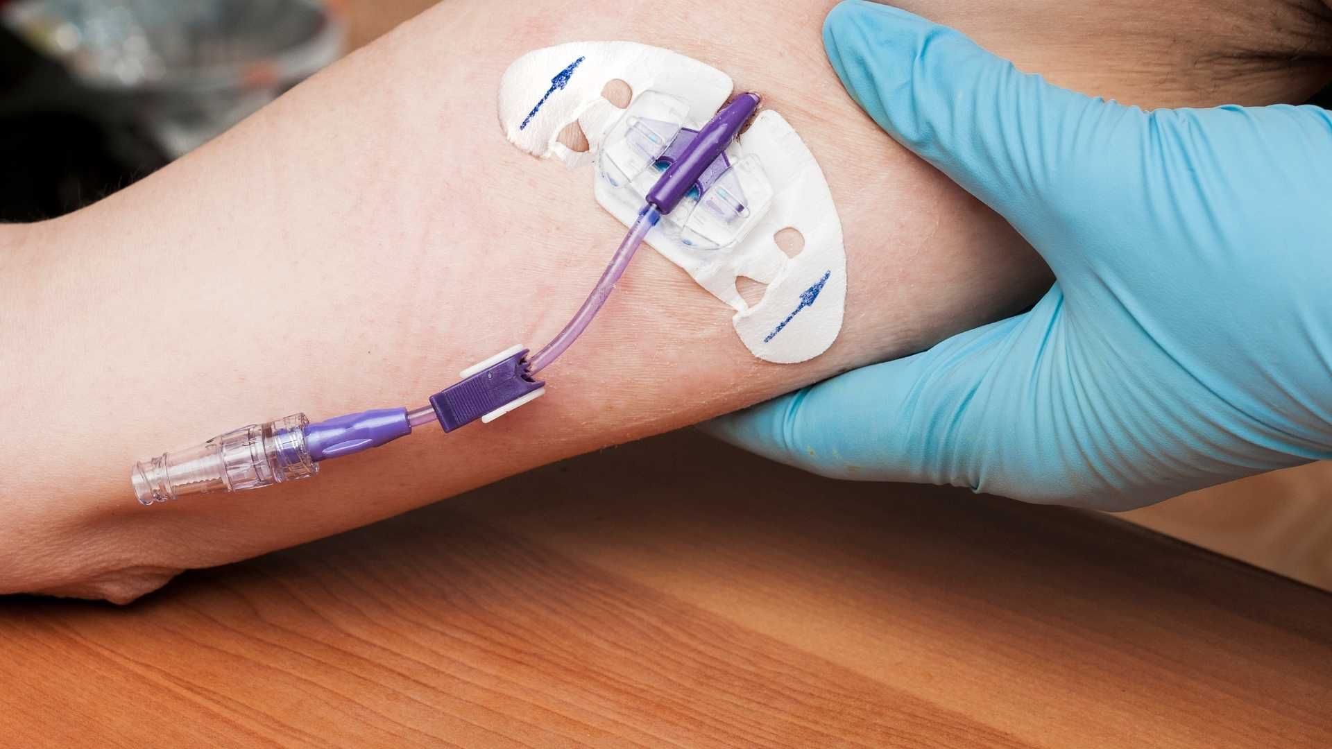
A Dual-Energy X-ray Absorptiometry (DXA) scan is considered the gold standard for analyzing body composition. It offers precise, detailed measurements of fat tissue, lean mass (which includes muscle), and bone mineral density. During the scan, a patient lies on a specialized table while a scanning arm passes over the body, taking images primarily of the hips, spine, and other key areas.
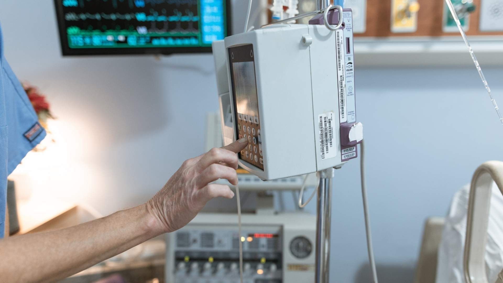
The process is quick, typically lasting about 10 to 20 minutes, and is non-invasive with minimal X-ray exposure, making it safe for repeated use. The resulting data provide an in-depth look at how fat and muscle are distributed throughout the body, helping to identify health risks and guide personalized health plans.
Metrics like Skeletal Muscle Mass (SMM) and Skeletal Muscle Mass Percentage (%SMM)
One of the key outputs of a DXA scan is the measurement of Skeletal Muscle Mass (SMM). This value indicates the total amount of muscle attached to the bones, which is vital for strength and mobility.
Along with SMM, the scan reports Skeletal Muscle Mass Percentage (%SMM), showing what fraction of the total lean mass is muscle. These metrics are crucial for assessing muscle health, especially in populations at risk for sarcopenia (age-related muscle loss) or muscle wasting due to illness.
Tracking these measurements over time enables health professionals and athletes to monitor muscle gains, losses, or stability, ensuring effective interventions and optimal performance.
The role of DXA in monitoring changes in muscle mass over time
For athletes and individuals undergoing weight management or rehabilitative programs, monitoring muscle mass is essential. DXA scans can be repeated periodically—every few months or annually—to observe progress.
Changes in Skeletal Muscle Mass and %SMM can influence nutritional and training strategies aimed at muscle preservation or growth. For example, post-scan consultations typically provide personalized advice on exercise routines, dietary adjustments, and other health interventions.
Additionally, DXA scans estimated resting metabolic rate (RMR), supporting tailored nutrition plans to maintain or increase muscle mass. Continuous monitoring allows for adjustments that optimize health outcomes, prevent muscle loss, and support overall vitality.
| Measurement | Purpose | Significance |
|---|---|---|
| Skeletal Muscle Mass | Total amount of muscle tissue | Guides strength and fitness planning |
| Skeletal Muscle % | Proportion of lean mass that is muscle | Assesses muscle health and aging |
| Body Fat Percentage | Distribution of fat tissue | Identifies health risks like visceral fat |
| Bone Density | Mineral content in bones | Evaluates osteoporosis risk |
Additional health insights from DXA scans
Besides muscle and fat analysis, DXA scans evaluate bone mineral density to assess fracture risk and bone health. The results are reported as T scores, comparing bone density to that of a healthy young adult.
Higher bone density scores indicate stronger bones, which reduce fracture risks. Regular DXA testing, especially for women over 50, helps monitor bone loss, facilitates early intervention, and supports the management of osteoporosis.
Understanding both muscle and bone health from DXA scans empowers individuals to take preventive measures and adopt lifestyle changes that enhance overall musculoskeletal health.
In summary, DXA scans provide comprehensive insights into body composition, outperforming traditional methods like calipers or bioelectrical impedance, and serve as an essential tool for personalized health and fitness management.
DXA Scans as a Tool for Monitoring and Supporting Muscle Mass
How do DXA scans help track muscle mass and body composition changes over time?
Dual-energy X-ray Absorptiometry (DXA) scans are highly effective in measuring body composition, including fat tissue, lean mass, and bone density. They provide detailed insights into the proportion and distribution of muscle, fat, and bone within the body. One of their primary uses is monitoring how muscle mass changes over periods, which is vital for athletes, older adults, and individuals undergoing rehabilitation.
By assessing metrics like Skeletal Muscle Mass (SMM) and Skeletal Muscle Mass Percentage (%SMM), DXA scans enable precise tracking of muscle development or loss. For example, measuring appendicular lean mass helps identify early signs of sarcopenia, or age-related muscle decline. These scans are especially useful for detecting subtle changes that might not be visible with other methods like BMI or bioelectrical impedance.
How does detailed analysis support fitness and health optimization?
The comprehensive analysis provided by DXA scans plays a significant role in personal health management. Athletes and fitness enthusiasts use these scans to tailor training programs by understanding their muscle and fat distribution accurately. This information helps optimize workouts, ensuring they target areas needing improvement while avoiding overtraining.
For general health, knowing one's body composition can highlight potential risks like excess visceral fat, which is linked to metabolic syndrome and cardiovascular disease. Regular monitoring fosters informed decisions about diet, exercise, and lifestyle adjustments, promoting better long-term health outcomes.
Post-scan consultations often include personalized advice based on the results. These may involve specific training recommendations, nutritional guidance, and strategies to preserve or increase muscle mass, especially as part of aging or in chronic health conditions.
Estimating Resting Metabolic Rate (RMR) to guide nutrition and exercise
DXA scans can provide an estimate of Resting Metabolic Rate (RMR), which reflects the number of calories the body needs at rest to maintain vital functions. Understanding RMR helps craft personalized nutrition plans that support muscle maintenance, fat loss, or gain.
By knowing the amount of lean mass, which is metabolically active, individuals can optimize their caloric intake and exercise routines. For instance, higher lean mass correlates with a higher RMR, indicating greater calorie burn even during rest.
This estimation allows for more effective diet and workout strategies, reducing guesswork in weight management and supporting muscle health over time. Whether for athletic performance, weight loss, or aging, leveraging RMR data enhances the precision of health interventions.
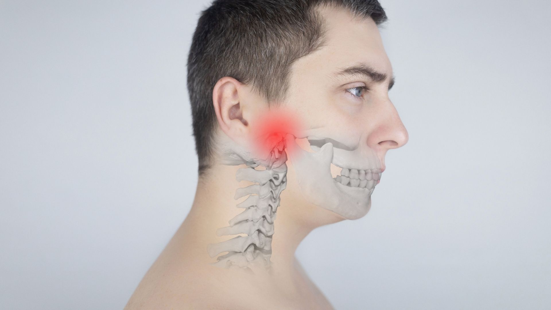
How do bone scans contribute to overall musculoskeletal health?
Bone health evaluation is another critical aspect of DXA scans. They measure bone mineral density, providing scores that help diagnose conditions such as osteoporosis and osteopenia. Bone scans involve lying on a special table while a scanning arm passes over the body, primarily targeting the hips and spine.
Results include interpretation of T scores, where higher scores suggest stronger bones, reducing fracture risk. Regular bone density assessments are essential, especially for postmenopausal women and older adults, as bone loss accelerates with age.
While bone scans focus on bones, their insights are indirectly related to muscle health. Stronger bones support better muscle function, and conditions like osteoporosis often coexist with muscle deterioration. Tracking bone density over time, along with muscle mass, offers a comprehensive view of musculoskeletal health.
The procedure and what it means for health monitoring
The DXA scan process is swift, painless, and involves low radiation exposure. Typically lasting about seven minutes, it uses special X-ray beams to measure body composition precisely.
Post-scan, detailed reports are generated, highlighting areas such as fat percentage, lean mass, and bone density. Healthcare providers interpret these results to develop individualized health or treatment plans.
By integrating this data, clinicians and individuals can identify potential health risks early, adapt lifestyle behaviors, and implement measures to maintain or improve musculoskeletal health.
Comparing DXA with other assessment methods for musculoskeletal health
Compared to other techniques like calipers or bioelectrical impedance, DXA scans offer more accurate and reliable measurements. They are regarded as the gold standard for body composition analysis.
While methods like BMI give only a rough estimate of body size, DXA provides detailed breakdowns of lean mass, fat distribution—including visceral fat—and bone quality.
This precision supports better diagnosis, monitoring, and intervention planning, which is especially critical for aging populations at risk of osteoporosis and muscle loss.
How body composition data can inform health decisions
Regular monitoring with DXA scans offers a means to track progress in fat loss, muscle gain, and bone health. This continuous assessment enables individuals to make informed decisions about diet, exercise, and lifestyle modifications.
For example, maintaining muscle mass through resistance training and proper nutrition can help counteract age-related sarcopenia, whereas monitoring bone density can prompt early treatment if decline is detected.
Conclusively, understanding body composition fosters proactive health management, improving quality of life and reducing the risk of chronic conditions.
| Aspect | Measurement Details | Health Implications |
|---|---|---|
| Muscle Mass (SMM, %SMM) | Quantifies skeletal muscle and its proportion | Detects muscle loss, guides fitness routines |
| Bone Density (T score) | Assesses mineral content and strength | Helps diagnose osteoporosis, fracture risk |
| Body Fat Percentage | Determines fat distribution, visceral fat | Risks for cardiovascular and metabolic diseases |
| Resting Metabolic Rate (RMR) | Estimates calorie needs based on lean mass | Guides nutrition and exercise planning |
Monitoring body composition with DXA scans thus offers a comprehensive picture of musculoskeletal health, vital for maintaining mobility, strength, and overall well-being.
Bone Scans in Diagnosing and Managing Musculoskeletal Conditions

What is the role of bone scans in detecting conditions like osteoporosis, fractures, infections, and tumors?
Bone scans are valuable diagnostic tools for uncovering problems within the skeletal system. They can detect early signs of osteoporosis, a condition where bones become fragile and more prone to fractures. By highlighting areas of decreased bone activity, bone scans help healthcare providers identify regions at higher risk.
In addition, bone scans are effective for identifying fractures that are not visible on standard X-rays, especially stress fractures or those hidden beneath other injuries.
Bone scans also play a crucial role in diagnosing infections such as osteomyelitis, by revealing increased bone activity where infection-induced inflammation occurs. Moreover, they assist in detecting bone tumors and metastases, showing abnormal areas of activity that suggest tumor growth or spread.
Overall, bone scans serve as a sensitive method to evaluate bone health and disorders, often revealing subtle or occult abnormalities that could be missed with other imaging techniques.
How do bone scans help monitor treatment responses and guide follow-up care?
Once a diagnosis is made, bone scans can be used to monitor the effectiveness of treatments for bone-related conditions. For example, in osteoporosis management, repeat scans help assess whether therapies like bisphosphonates are strengthening bones.
In cases of infections, follow-up scans can confirm whether the infection has resolved or if there is ongoing activity requiring further treatment.
Similarly, for patients treated for bone tumors or metastases, serial scans are invaluable for tracking disease progression or regression, allowing clinicians to adjust treatment plans accordingly.
This ongoing evaluation supports personalized healthcare strategies, ensuring that interventions remain effective over time and reducing the risk of fractures or other complications.
How do bone scans complement other imaging methods like MRI and CT?
While bone scans excel at detecting metabolic activity and early abnormalities in the bones, they are often used in conjunction with other imaging techniques for comprehensive assessment.
Magnetic Resonance Imaging (MRI) offers superior soft tissue contrast, making it preferable for evaluating muscles, ligaments, cartilage, and nerve involvement. MRI is particularly useful when soft tissue injury is suspected or detailed anatomical visualization is needed.
Computed Tomography (CT) scans provide high-resolution images of bone structure and are excellent for assessing complex fractures, bone geometry, and detailed tumor characterization.
Combining the functional information from bone scans with the anatomical details from MRI and CT makes it possible to achieve a precise diagnosis and tailor treatment strategies. For example, a suspected bone tumor might be imaged with CT for structural assessment, MRI for soft tissue involvement, and bone scan to evaluate metabolic activity or metastasis.
In conclusion, these imaging modalities work together to provide a comprehensive picture of musculoskeletal health, allowing for more accurate diagnosis, better treatment planning, and effective follow-up.
| Imaging Modality | Strengths | Typical Use Cases | Additional Notes |
|---|---|---|---|
| Bone Scans | Detect metabolic activity, early bone changes | Osteoporosis, infections, fractures, tumors | Sensitive to changes before structural abnormalities appear |
| MRI | Soft tissue detail, nerve and cartilage visualization | Soft tissue injuries, nerve impingement, tumor extent | No radiation, longer scan times |
| CT | Detailed bone anatomy, complex fractures | Fracture evaluation, tumor visualization | Higher radiation exposure |
| Integration | Combining scans increases accuracy and detail | Comprehensive musculoskeletal assessment | Improves diagnosis and personalized treatment planning |
Understanding and utilizing these tools effectively help healthcare providers monitor bone and muscle health, manage existing conditions, and prevent future problems.
Assessing Body Composition and its Role in Musculoskeletal Health

Why is body composition analysis important for overall health?
Understanding body composition is vital because it offers detailed insights into the distribution of fat, lean muscle, and bone mass in the body. Unlike BMI, which provides a simple weight-to-height ratio, methods like DEXA scans can accurately measure these components. This detailed information helps identify potential health risks such as excess visceral fat—which is linked to heart disease and diabetes—and muscle loss, which can lead to decreased strength and mobility.
Accurate body composition analysis also aids in personalizing fitness and nutrition strategies. For example, it allows individuals to tailor their exercise programs to focus on either fat loss or muscle building, depending on their specific needs.
How does a DEXA scan compare with other methods?
The DEXA scan is considered the gold standard for assessing body composition because of its high accuracy and reliability. It provides a three-compartment analysis, breaking down fat mass, lean tissue (including muscle), and bone density.
Compared to other techniques:
| Method | Precision | Speed | Cost | Additional Insights |
|---|---|---|---|---|
| Bioelectrical Impedance | Moderate | Fast | Low | Less reliable, affected by hydration |
| Skinfold Calipers | Moderate | Slow | Low | Operator-dependent, less comprehensive |
| Hydrostatic Weighing | High | Slow | Moderate | Requires water submersion |
| Bod Pod (Plethysmography) | High | Fast | Moderate | Equipment costly |
| DEXA Scan | Very High | Quick (10–20 min) | Higher | Offers detailed skeletal and soft tissue data |
This detailed approach makes DEXA ideal for routine monitoring and clinical evaluation.
How do scans help identify health risks like osteoporosis and sarcopenia?
Bone health is a crucial part of musculoskeletal health. DXA scans measure bone mineral density precisely, reporting results as T scores. Scores closer to zero indicate stronger, healthier bones. Lower scores signal osteoporosis or osteopenia, conditions that increase fracture risk.
Monitoring bone density over time can detect early bone loss, allowing timely treatment. Measures such as calcium and vitamin D supplementation, medications to slow bone loss, or lifestyle changes like weight-bearing exercise can be recommended to maintain or improve bone health.
Similarly, DXA scans help spot sarcopenia, characterized by loss of muscle strength and mass. Recognizing sarcopenia early grants opportunities for intervention through resistance training and nutritional support, which help preserve muscle function and independence.
How does the connection between bone health assessments and muscle preservation influence health strategies?
Bone and muscle tissues are closely connected, both physically and biologically. The health of one strongly influences the other. For instance, loss in bone density and muscle mass often occur simultaneously, especially in aging populations.
By evaluating both tissues with DXA scans, healthcare providers can tailor strategies that address both issues. Combining resistance exercises to build muscle and improve bone density, along with dietary adjustments rich in calcium and vitamin D, forms an effective approach to maintaining musculoskeletal integrity.
Research shows that improving muscle strength can reduce fracture risk by supporting overall stability and reducing fall likelihood. Conversely, stronger bones can support richer muscle activity.
How does regular monitoring impact long-term health management?
Consistent body composition assessments enable the early detection of undesirable changes, such as muscle wasting or bone loss. This proactive approach supports preventive care, reducing the likelihood of fractures, mobility issues, and chronic diseases.
Regular DEXA scans motivate individuals to stick with healthy lifestyle habits, providing tangible progress reports. These results inform healthcare providers’ decisions, allowing tailored interventions—such as adjusting exercise routines or revising nutritional plans.
Ultimately, continuous monitoring fosters a preventive framework that promotes healthier aging, enhances quality of life, and reduces healthcare costs.
| Benefits of Regular Body Composition Monitoring | Practical Applications | Long-term Impacts |
|---|---|---|
| Early detection of sarcopenia and osteoporosis | Personalized workout and diet plans | Better mobility and reduced fracture risk |
| Tracking changes over time | Adjusting treatments as needed | Slower biological aging processes |
| Motivation for lifestyle improvements | Motivation through progress reports | Enhanced overall well-being |
Gaining precise and timely insights through advanced scans like DEXA is integral to holistic health management, supporting both preventative and therapeutic goals.
The Future of Bone Scans in Supporting Aging Populations
What are the recent developments in bone scan technology regarding muscle and bone health assessment?
Recent advances in bone scan technology have significantly enhanced our ability to evaluate both bone and muscle health with greater precision and safety. New imaging methods, such as high-resolution DXA and enhanced software algorithms, allow clinicians to detect even subtle changes in bone density and muscle mass. These improvements facilitate earlier diagnosis of conditions like osteoporosis, sarcopenia, and other degenerative musculoskeletal disorders.
Integration with other diagnostic tools—such as biomarkers, MRI, and CT scans—further refines the accuracy of assessments. This multi-modal approach offers a comprehensive view of musculoskeletal health, leading to more tailored interventions.
In space medicine, studies modeling microgravity effects reveal how bones and muscles adapt and deteriorate in low gravity environments. These insights, aided by advanced imaging like DXA, help in developing effective countermeasures to prevent bone density loss and muscle atrophy, both critical for astronauts and aging individuals alike.
These innovations are paving the way for personalized health management strategies. Early detection and intervention, powered by improved imaging, can dramatically improve quality of life for older adults by delaying or preventing severe musculoskeletal decline.
How is research on microgravity contributing to overall musculoskeletal health understanding?
Microgravity research offers unique insights into bone and muscle biology by mimicking accelerated aging processes. When subjected to space conditions, bones lose approximately 1% of their density each month without countermeasures, similar to osteoporosis development on Earth.
Scans such as DXA are crucial in these studies because they monitor changes in bone mass and composition, providing data to test interventions like specialized exercise routines and pharmaceutical treatments.
Understanding how tissues reshape in low gravity environments helps scientists design better strategies for maintaining skeletal integrity here on Earth. For instance, treatments like myostatin inhibitors are being explored to prevent muscle wasting, inspired by microgravity studies.
Furthermore, tissue chips and other laboratory models, combined with imaging insights, drive advances in countermeasures, which benefit both space explorers and aging populations prone to osteoporosis and muscle loss.
What is the potential for tracking muscle and bone health in elderly populations?
The potential for utilizing advanced bone scans in elderly health management is immense. Regular assessments can help detect early signs of osteoporosis and sarcopenia, conditions that significantly increase fracture risk and mobility issues.
By monitoring changes over time, clinicians can tailor nutrition, exercise, and medication plans to individual needs, thereby enhancing treatment efficacy.
Innovative imaging techniques allow for precise measurement of visceral fat, which is linked to metabolic syndromes such as diabetes and cardiovascular disease. This comprehensive approach supports proactive health management.
Furthermore, incorporating these scans into routine check-ups enables healthcare providers to identify risks before symptoms become severe, encouraging preventative measures.
Advancements in scan technology and interpretation
The evolution of scan technology focuses on enhanced clarity and lower radiation exposure. AI-driven interpretation software helps in quickly analyzing results, identifying early signs of disease, and predicting progression.
This technological progress supports large-scale screening programs, especially in aging populations where early intervention can prevent serious health crises.
Microgravity research and space medicine
Applying space medicine principles, especially the study of tissue adaptations in microgravity, informs the development of innovative therapies for age-related decline. Portable, precise imaging devices like updated DXA machines enable in-flight monitoring of astronauts’ bone and muscle health, which could inspire similar applications on Earth.
Potential for tracking muscle and bone health in elderly populations
Routine use of advanced scans in geriatrics offers a pathway to personalized medicine, allowing tailored interventions aimed at preserving mobility and independence.
| Aspect | Description | Practical Implication |
|---|---|---|
| Imaging Precision | Enhanced resolution and software analysis | Detect early tissue changes |
| Safety | Reduced radiation doses | Safe for regular use in vulnerable populations |
| Integration | Combining imaging with biomarkers and AI | Better diagnostic accuracy and predictive analytics |
| Space Research Insights | Understanding tissue plasticity and adaptation in microgravity | Developing new treatments for age-related decline |
| Preventative Measures | Early detection leading to timely intervention | Slowing down or reversing musculoskeletal deterioration |
This continual evolution in bone and muscle imaging technology promises to significantly improve health management for aging populations, helping them maintain independence, prevent fractures, and enjoy a higher quality of life.
Concluding Insights: The Power of Bone Scans in Maintaining Musculoskeletal Health
Bone scans, especially DXA, have revolutionized our approach to musculoskeletal health by providing detailed, accurate insights into bone and muscle status. They not only facilitate early detection of conditions like osteoporosis and sarcopenia but also support ongoing monitoring, helping individuals and healthcare providers craft personalized strategies for maintaining strength and mobility. As technology advances, bone scans will become even more integral in comprehensive health assessments, including in innovative fields like space medicine. Embracing these diagnostic tools ensures a proactive stance in preserving muscle mass and overall musculoskeletal function—crucial for healthy aging and enhanced quality of life.
References
- DXA body composition analysis | Sports Medicine - UC Davis Health
- DXA Scan (Bone Density Test): What Is It & How It's Done
- Understanding Body Composition: How DEXA Scans Can Optimize ...
- Bone scan - Mayo Clinic
- DXA Body Composition Analysis - Sports Medicine - UC Davis Health
- The Importance of Monitoring Body Composition and Muscle Mass
- Bone scan | Tests and scans - Cancer Research UK
- Nuclear Medicine Musculoskeletal Assessment, Protocols ... - NCBI
- The Benefits of a DEXA Body Composition Scan - Exercise Matters
- Triple Phase Bone Scan - StatPearls - NCBI Bookshelf